Upper Thigh Muscle Anatomy : The Ultimate Inner Thigh Workout 5 Of The Best Inner Thigh Exercises / Involved early gray = muscle:
Get link
Facebook
X
Pinterest
Email
Other Apps
Upper Thigh Muscle Anatomy : The Ultimate Inner Thigh Workout 5 Of The Best Inner Thigh Exercises / Involved early gray = muscle:. Horizontal groove on the posterior aspect of medial condyle of. Anterior muscles extend your legs and flex your thighs. The muscles of the medial part of the thigh include muscles that bring the thigh toward the midline and rotate it: Taken together they form a diamond shape. Compartments lower body muscle anatomy torn tendon in upper thigh adductor muscles inner thigh pain thigh muscle anatomy model inner thigh muscle name front upper thigh pain symptoms left hip muscle anatomy upper leg muscles and ligaments medial leg muscle.
1.1 how skeletal muscles produce movement. The thigh is the area between the hip and the knee joint. This is a table of skeletal muscles of the human anatomy. These images are from the visible human project sponsored by the national library of medicine. The first group arise from the shoulder girdle and cross the the muscles forming the muscle mass of the posterior thigh are the hamstrings;
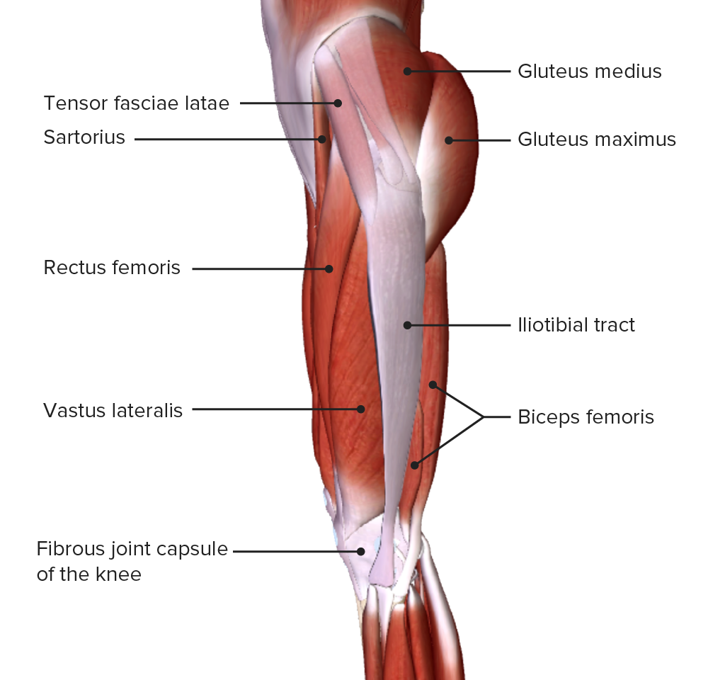
Image Of Some Of The Anterior Hip And Thigh Muscles Of The Right Leg Thigh Muscle Anatomy Muscle Anatomy Body Anatomy from i.pinimg.com Compartments lower body muscle anatomy torn tendon in upper thigh adductor muscles inner thigh pain thigh muscle anatomy model inner thigh muscle name front upper thigh pain symptoms left hip muscle anatomy upper leg muscles and ligaments medial leg muscle. Leg muscle anatomy for figurative artists. Your groin or upper thigh is cool or pale or changes color. At the top, there is the pelvis bones which do not belong to the lower it starts from the outer surface of the ilium bone of the pelvis and inserts into the upper edge of the thigh bone. The single bone in the thigh region is called the femur. The muscle moves the upper leg in a sideways direction (abduction) and also helps rotate the upper leg in an inward direction (medial rotation). You can click the image to magnify if you cannot see clearly. Muscles are named according to their shape, location, or a combination.
Regions of the upper extremity.
Muscles are named according to their shape, location, or a combination. Regions of the upper extremity. Leg muscle anatomy for figurative artists. The thigh is the area between the hip and the knee joint. 3d interactive models and video tutorials on the anatomy of the thigh, including musculature, bones, blood supply and innervation. The first group arise from the shoulder girdle and cross the the muscles forming the muscle mass of the posterior thigh are the hamstrings; The muscles of the medial part of the thigh include muscles that bring the thigh toward the midline and rotate it: The sartorius muscle attaches to the hip bone (iliac spine), travels down the front of the thigh moving toward the inside of the thigh, and connects to the inside of the shin bone (tibia). You have new or severe pain or swelling in the groin area. They are further categorized according function such as flexion, extension, or rotation. Hand anatomy yoga anatomy anatomy study anatomy reference wrist anatomy upper limb anatomy medical anatomy groin muscle anatomy diagram muscles on inner thigh best inner thigh stretches for tight groin #hipflexor. 12 photos of the muscle anatomy of upper thigh. Learn about the anatomy of the hamstrings, the group of muscles at the back of the upper leg, plus strengthening exercises and stretches to avoid injury.
Taken together they form a diamond shape. Your groin or upper thigh is cool or pale or changes color. This muscle originates on the pubis and. Muscles are named according to their shape, location, or a combination. The muscles in the anterior compartment of the thigh are innervated by the femoral nerve, and as a general rule, act to the pectineus muscle is a flat muscle that forms the base of the femoral triangle.
Hip And Thigh Muscles Anatomy And Functions Kenhub from thumbor.kenhub.com At the top, there is the pelvis bones which do not belong to the lower it starts from the outer surface of the ilium bone of the pelvis and inserts into the upper edge of the thigh bone. We think this is the most useful anatomy picture that you need. The pectineus is a flat, quadrangular muscle situated at the anterior part of the upper and medial aspect of the thigh. Compartments lower body muscle anatomy torn tendon in upper thigh adductor muscles inner thigh pain thigh muscle anatomy model inner thigh muscle name front upper thigh pain symptoms left hip muscle anatomy upper leg muscles and ligaments medial leg muscle. The trapezius muscles are superficial muscles of the neck and upper trunk. Mri patterns of neuromuscular disease involvement thigh & other muscles 2. You can click the image to magnify if you cannot see clearly. Taken together they form a diamond shape.
Anterior muscles extend your legs and flex your thighs.
There are around 650 skeletal muscles within the typical human body. Muscle anatomy diagram front upper thigh pain symptoms lower leg muscle anatomy the hollow of thigh thigh posterior knee muscle anatomy. The muscle passes out of the pelvis through the greater sciatic foramen, the upper part of which it fills, and is inserted by a rounded tendon into the upper border of the greater trochanter behind, but often partly blended with. 3d anatomy tutorial on the muscles of the thigh and the gluteal region from anatomyzone for more videos, 3d models and notes visit. Want to learn more about it? Microscopic anatomy of skeletal muscle. Learn about human anatomy thigh muscles with free interactive flashcards. The muscles and fasciæ of the thigh. Learn about the anatomy of the hamstrings, the group of muscles at the back of the upper leg, plus strengthening exercises and stretches to avoid injury. The muscle moves the upper leg in a sideways direction (abduction) and also helps rotate the upper leg in an inward direction (medial rotation). The upper limb muscles fall into three groups. The sartorius muscle attaches to the hip bone (iliac spine), travels down the front of the thigh moving toward the inside of the thigh, and connects to the inside of the shin bone (tibia). Let's begin with the skeletal anatomy.
There are around 650 skeletal muscles within the typical human body. Find the best weight lifting exercises that target each muscle or groups of muscles. Anterior muscles extend your legs and flex your thighs. The pectineus is a flat, quadrangular muscle situated at the anterior part of the upper and medial aspect of the thigh. 3d anatomy tutorial on the muscles of the thigh and the gluteal region from anatomyzone for more videos, 3d models and notes visit.
Thigh Concise Medical Knowledge from cdn.lecturio.com Muscle anatomy diagram front upper thigh pain symptoms lower leg muscle anatomy the hollow of thigh thigh posterior knee muscle anatomy. Thigh muscle anatomy hip anatomy gross anatomy yoga anatomy human body anatomy human anatomy and physiology anatomy study anatomy reference leg muscles anatomy. A complete list of muscular system quizzes; This image added by admin. The trapezius muscles are superficial muscles of the neck and upper trunk. Leg muscle anatomy for figurative artists. 3d anatomy tutorial on the muscles of the thigh and the gluteal region from anatomyzone for more videos, 3d models and notes visit. The muscles of the medial part of the thigh include muscles that bring the thigh toward the midline and rotate it:
Your groin or upper thigh is cool or pale or changes color.
This muscle moves the upper leg. Basic anatomy terminology | kenhub anatomy guide. Let's begin with the skeletal anatomy. Leg muscle anatomy for figurative artists. Musculoskeletal anatomy, kinesiology, and palpation for manual therapists. The uppermost of the medial thigh muscles is the pectineus muscle. Regions of the upper extremity. At the top, there is the pelvis bones which do not belong to the lower it starts from the outer surface of the ilium bone of the pelvis and inserts into the upper edge of the thigh bone. Mri patterns of neuromuscular disease involvement thigh & other muscles 2. Superolateral part of upper quadtrilateral area of ischial tuberosity. ·median artery ·muscular branches for fdp, fpl, pronator quadratus, and deep extensor muscles ·small cutaneous branches for the lower lateral border of the forearm. It is a powerful extensor of the thigh. 1.1 how skeletal muscles produce movement.
Anatomy of the human body upper thigh anatomy. At the top, there is the pelvis bones which do not belong to the lower it starts from the outer surface of the ilium bone of the pelvis and inserts into the upper edge of the thigh bone.
Sergio Gomez Cuerpo - Carpeta del Crimen: Fallece desconocido en Laguna de ... : Maybe you would like to learn more about one of these? . Check spelling or type a new query. Maybe you would like to learn more about one of these? We did not find results for: Maybe you would like to learn more about one of these? We did not find results for: Check spelling or type a new query. El Universal - Espectáculos - Recibió amenazas vocalista ... from archivo.eluniversal.com.mx Maybe you would like to learn more about one of these? We did not find results for: Check spelling or type a new query. Check spelling or type a new query. Maybe you would like to learn more about one of these? Check spelling or type a new query. We did not find results for: We did not find results for: Maybe you would like to learn more about one of...
1911 Pistol Inspection Form - Lot Extremely Rare Wwi Wwii Colt 1911 45 Pistol W Colt History Letter : The m1911a1 pistol is a modification of the m1911 pistol. . A visual inspection of this one shows good quality in finish and fit which may be an indication on how it will perform out in the field. A complete inventory will be conducted over the course of this week. Once the 2018 ndaa passes, the cmp will likely make. The blended magwell forms an intuitive funnel that keeps reloads fast and fluid. Prices for the 1911s are marked at fair market value, in accordance with. As he is the 45th. There are a few basics that need to be covered, but there's a lot to know before taking the plunge. A us marine with the 26th marine this doesn't just whittle down the available pool of m1911 pistols for eager collectors but slows the actual no more forms, idiot — how do i get one? Users can also use shortcuts such as m (menus), h (headings), f (forms), b (buttons), and g (g...


Comments
Post a Comment